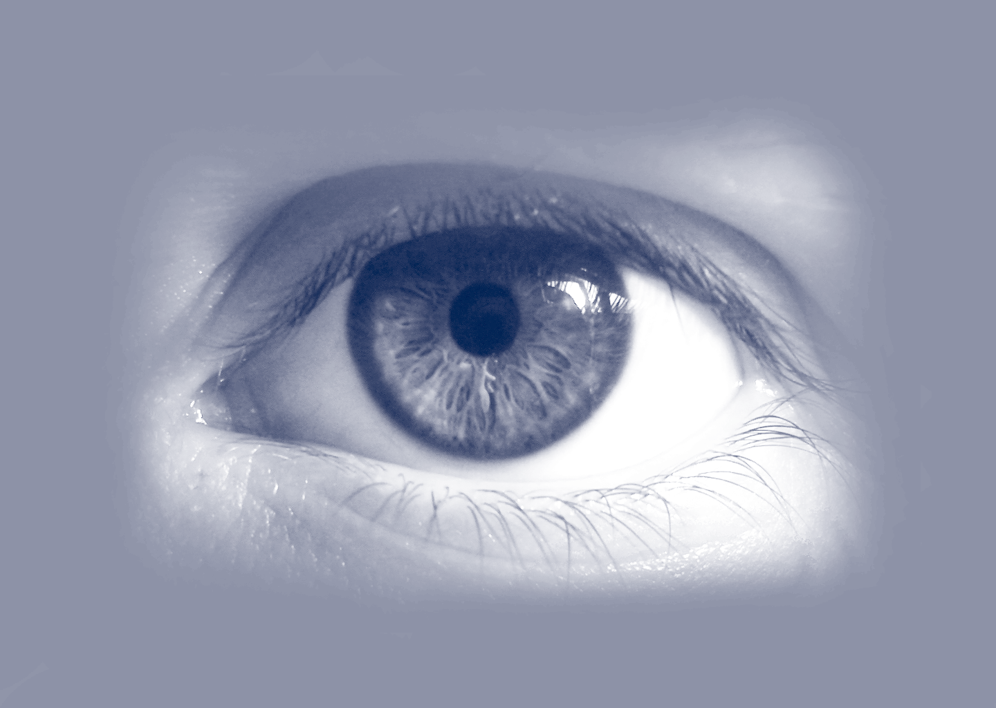
close up of eye

Lumedica Vision

MANAGEMENT: Adam Wax
DUKE INVENTOR: Adam Wax
lumedicavision.com

Lumedica Vision was founded by an experienced team of engineers including Chief Scientist Dr. Adam Wax from the Pratt School of Engineering. Dr. Wax’s team has developed a new low-cost optical coherence tomography (OCT) scanner that could dramatically extend the impact of the imaging technology for eye health by making eye imaging more affordable, accessible and easier to use. Affordable access would enable more health care providers to conduct OCT imaging, giving patients greater, earlier access to the one test that can save their vision and improve their quality of life.

Lumedica Vision Logo
Lumedica OCT Prototype

THE PROBLEM:

Seen as the gold standard for early detection of diseases including macular degeneration, diabetic retinopathy and glaucoma – each of which can threaten vision while remaining symptomless – OCT remains an expensive procedure.  “Once you have lost vision, it’s very difficult to get it back, so the key to preventing blindness is early detection,” said Adam Wax

The challenge is access.

  • Current OCT focus on features keeps prices high and out of reach
  • Many developing countries have little or no access to OCT
  • Older OCTs in Eastern Europe have limited diagnostic efficacy
  • US Optometrists: 40% balk at high price of entry
  • Primary Care Physicians: Too complex and expensive to operate

THE SOLUTION:

“Our goal is to make OCT drastically less expensive, so more clinics can afford the devices, especially in global health settings,” said Wax

The key to the cost reduction is the innovative spectrometer design. This approach takes light on a circular path – rather than the conventional combination of lenses, mirrors and precision diffractive optics. The overall result is a product which reduces the typical weight of a clinical OCT system from 50-70 pounds down to just 4 pounds, while cutting the cost from at least $60,000 to less than $14,000.
What is OCT?

Optical Coherence Tomography is the light-equivalent to SONAR, sending signals to, then reflecting off of surfaces in order to measure depths of individual layers. Those collective signal measurements form an image of the retinal layers, where eye diseases quietly lurk, and can be accurately detected. Although OCT has been available for over 25 years, it remains an expensive technology to manufacture and operate. The use of high-quality optics, precision components and complex software have put this technology out of reach for most healthcare providers worldwide.

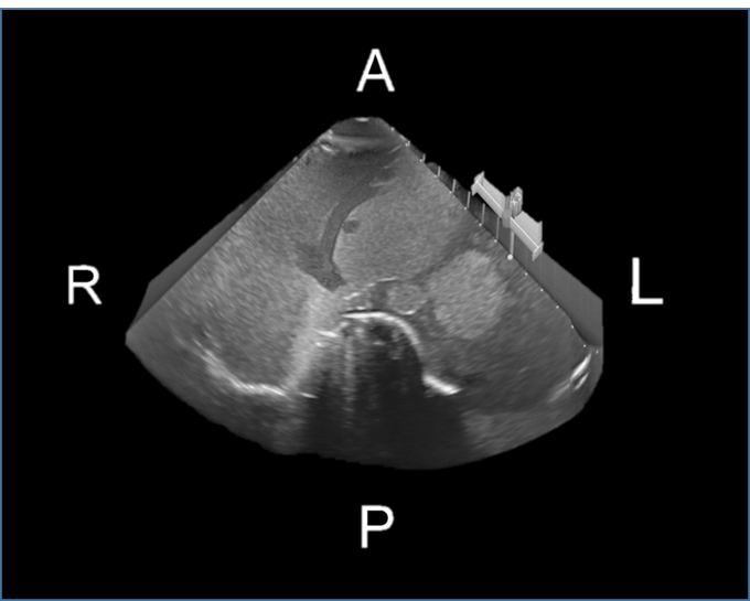
ultrasound image

Omnisono

DUKE INVENTOR: Josh Broder

Duke/Stanford spinout OmniSono has developed a new low-cost system enabling 2D ultrasound machines to perform rapid, high-quality, oriented 3D and multiplanar ultrasound imaging. With a price-point less than 1/10th that of existing 3D solutions and features for ease-of-use, OmniSono will greatly expand the accessibility and size of the ultrasound market.

Omnisono logo
lindy microglassification

Lindy Biosciences

MANAGEMENT: Deborah Bitterfield
DUKE INVENTOR: David Needham
lindybio.com

Lindy Biosciences is a development-stage protein therapeutic formulations company. Our core technology produces spherical, dense, stable particles of a therapeutic protein. These protein particles are ideal for long-term storage, or for non-standard formulations such as suspension formulations (for high-concentration delivery of antibodies), encapsulation (for sustained/controlled release), or dry powder pulmonary delivery.​

lindy biosciences logo

MICROGLASSIFICATION™

Produces dense, dry, stable protein particles ideal for suspensions or encapsulation

Microglassification™ is a process that gently removes a majority of the water from solutions of proteins, or other biologics, resulting in solid, spherical, amorphous microbeads. In this dry state, biologics are often stable enough for long-term storage, transport, or incorporation into drug delivery formulations.

The video shows one of our early experiments: Microglassification™ of a single protein microdroplet, held on the end of a glass capillary micropipette. The resulting solid microbead consists of pure protein at > 1 g/mL. Currently used methods mimic this experiment, producing protein microdroplets on a bulk scale and dehydrating those microdroplets within seconds.

High-Concentration Biologics

Biological molecules such as monoclonal antibodies often require dosing of several hundred mg. In order to allow subcutaneous administration, which can only accomodate 1-2 mL of volume, the protein must be formulated at high concentration (>200 mg/mL). However, the viscosity of solutions of these molecules increases dramatically above 100 mg/mL, making injection prohibitively difficult.

Suspensions of Microglassified protein can have a much lower viscosity than solutions of the same concentration. Preliminary formulations have been injectable through a 27G needle at >500 mg/mL.

Metacept Systems

MANAGEMENT:
DUKE INVENTOR: David Smith
metaceptsystems.com

Metacept provides design and engineering services for complete electromagnetic systems, with extensive experience in transitioning metamaterials to real-world applications. We can provide end-to-end development resources.

metacept logo

THE TECHNOLOGY

Imaging

Rethinking the problem of electromagnetic imaging from the ground up, Metacept designs hardware together with software to reach the physical limits of imaging technology.

Metamaterials

Metamaterials are a novel way to engineer material properties using micro-structural design. Metamaterials are able to intricately and precisely control many types of wave dynamics from electromagnetic to acoustic fields.

Physics-based Deep Learning

Deep learning has enabled machines to perform classification tasks. Metacept goes a step further to physically implement neural networks with electromagnetic waves to process data at the speed of light.

doctor holding tablet with manage my surgery app on display

Higgs Bosun Health

MANAGEMENT: Rajeev Dharmapurikar
DUKE INVENTORS: Nandan Lad, Ziad Gellad
higgsbosonhealth.com

Manage My Surgery (MMS) is developed by Higgs Boson, LLC and is a revolutionary application which navigates the patient through all phases of their surgical procedure. It provides a secure and HIPAA-compliant mobile platform which fosters communication between the patient, practice, and care provides. We have created different modules in Gastroenterology, Neuromodulation and Spine.

Manage my surgery logo

An overwhelming body of evidence has shown that patients who are more actively engaged in their care experience better health outcomes and incur lower costs. Preparation for an upcoming procedure can be a stressful process for a patient. Can I eat 24 hours prior? When do I stop my medications? How early do I need to arrive? Can I drive home? What type of pain can I expect post-procedure? Manage My Surgery provides real-time answers to these questions and more via a robust and user-friendly tool which empowers the patient throughout the life-cycle of the procedure. It provides patients, family members and providers reminders, checklists and procedure-specific information on one integrated mobile platform.

Higgs Boson, LLC consists of a team of seasoned physicians, surgeons (including 2 Duke faculty members) and information technology professionals who believe that the right data, in the right hands, at the right time can improve patient satisfaction and provide better outcomes.

Mobile Application

Prepped is a mobile application that generates helpful, timely suggested questions for patients and caregivers to ask their clinical providers. The app uses a strength in numbers in approach – Prepped crowdsources questions submitted by national experts, patients, caregivers and other users to recommend the best questions for the patient based on the patient’s profile and where they are in the disease process. Current versions of Prepped are focused on patients with cancer and/or COPD

Female doctor advising patient in hospital office during regular medical exam, healthcare and prevention concept.

MeTree

MANAGEMENT: Lori Orlando
DUKE INVENTOR: Geoff Ginsburg

MeTree is a family health history tool. Prior to an appointment, the doctor’s office will send an app-based survey for your/your family health history. Based on this data, the product will run algorithms, generate a report, and provide recommendations on lifestyle interventions with an overall goal of building a database of health, diseases, and outcomes.

metree logo

Family history in primary care

The systematic collection of family health history (FHH) can identify individuals at increased risk for common diseases including cardiovascular disease, diabetes, and cancer; many evidence-based guidelines rely upon risk stratification using FHH to guide the appropriate use of alternative (non-routine) screening procedures (such as breast MRI), diagnostic tests, and/or genetic counseling.

Unfortunately there are several barriers to the adequate collection of FHH within routine primary care: lack of patient preparation to provide FHH, the amount of time needed to collect FHH, lack of standardization, and limited training in synthesizing FHH data into a clinically actionable care plan.

MeTree

To address these issues, we have created a web-based patient-entered risk stratification and clinical decision support tool, MeTree, with the following design goals:

  • Develop a collection interface that is easy for patients to use and facilitates collection of all the necessary FHH and personal history components to perform risk stratification (full 3-generation pedigree with age of disease onset, current age or age of death, and cause of death for each relative)
  • Provide lay level and technical decision support that is clinically actionable for providers, activating for patients, and easy for patients and providers to understand • Base decision support upon guidelines widely accepted by PCPs and provide just-in-time education within the reports about what criteria triggered the recommendation, points to consider about the recommendation, data such as NNT, references to the guidelines, and links to additional resources
  • Capitalize on the patient-provider encounter to encourage discussions of preventive health and disease risk management.

MeTree will be available as an independent web-service that provides risk stratification and clinical decision support for the following conditions: breast cancer, ovarian cancer, colon cancer, hereditary cancer syndromes, coronary artery disease, hereditary cardiovascular conditions, stroke, aortic aneurysm, diabetes, and hereditary liver diseases (such as hemochromatosis).

security camera

Airgus

MANAGEMENT: Daniel Reichman
DUKE INVENTOR: Jordan Malof, Leslie Collins
ai-rgus.com

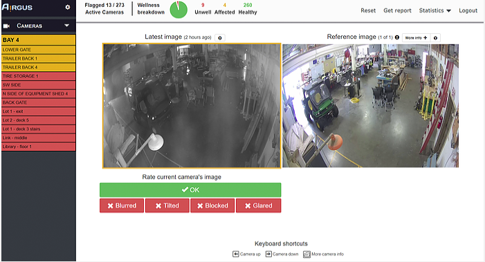
AᴉRGUS is an AI-based, low-cost, easy to use, and highly accurate solution. AᴉRGUS automatically inspects each camera and alerts you when the view is compromised by:

▪ Blur ▪ Tilt ▪ Tamper ▪ Obstruction ▪ Glare ▪ Black/blank screen ▪ More on request

ai-rgus logo

AᴉRGUS Dashboard

AᴉRGUS’s simple user interface saves operators’ time by streamlining the camera inspection process. You will also get reports, alerts, statistics and even work orders to make correcting your cameras’ wellness easy.

airgus dashboard
doctor holding clipboard with 3d heart overlay

Heart View Medical APS

MANAGEMENT:
DUKE INVENTOR: Olaf von Ramm
heartviewmedical.com

Our vision is to redefine the cardiology community’s understanding of the human heartbeat. HeartView Medical is developing the next generation high speed ultrasound system with extensive algorithms that enable automated quantitative measurements of the heart in a 3D view within one heart beat compared to current technology.

Heart View Medical Logo

ABOUT

 
Current state of the art: While imaging techniques are central to these advances, limitations exist in current imaging methods in their ability to rapidly record cardiac events (at physiologic speeds). At present, recording speeds are insufficient to properly link electrical activation to mechanical events and flow. ECG record activity in the 1-2 millisecond, i.e. 500-1000 events per second range while imaging methods (angiography, computed tomography-CT, magnetic resonance imaging-MRI and echocardiography) can record events between 5 to 100 times per second, thus substantially slower than ECG. In the case of 3D echo, CT and MRI, the problem is further confounded by the necessity to create images over multiple heartbeats where only portions of the image are obtained in a single heartbeat.A breakthrough in imaging of the heart at very high acquisition rates (physiologic rate imaging) is needed to further advance our understanding of the relationship of electrical, mechanical and flow events. Because of current slow imaging rates, it is likely that certain cardiac abnormalities remain undetectable. Developing a method to link these various cardiac activities holds a promise of providing a much more detailed understanding of how the heart works in normal and abnormal conditions with a potential to detect heart diseases earlier and to evaluate the results of current therapies more precisely and understand the impact of major risk factors such as diabetes and hypertension on the myocardium to open for new therapies. Moreover, echocardiography would improve the understanding of arrhythmia development for electrophysiology procedures and shorten procedure time and minimize patient risk.

SOLUTION

 
With a high-speed echocardiographic system Heart will change the way a heartbeat is understood from a diagnostic perspective. This means:
  • Diagnostic improvement for heart failure and tailored therapy with heart failure devices
  • Improved arrhythmia detection
  • Increased capability in detection of hypertrophy, fibrosis and scar
  • Potential to identify subclinical heart disease
  • Reduced need for MRI and CT
  • Substantially reduced acquisition time.
Gavilan scientist playing chess with green virus

Gavilán Biodesign

MANAGEMENT:
DUKE INVENTOR: Bruce Donald, Jonathan Jou, Marcel Frenkel, Mark Hallen
gavilanbio.com

Gavilán Biodesign combines state of the art physics-based modeling with our unique high accuracy AI platform to computationally screen trillions of molecules in order to find therapeutics that can overcome resistance. Gavilán is the first to add the dimension of evolution and time in our screens! We do this through our proprietary design software suite called Sylph that can design both biologics and small molecules for specificity and resistance resilience.   

gavilan logo

Our algorithms are based on rigorous mathematical guarantees allowing us to make confident predictions about our chemical space and continuously improve our predictions through improvements to our entropy-aware, continuously flexible, high accuracy biophysical models that are already the most sophisticated models of their kind for combinatorial drug design.

Explore Beyond, Leave Nothing Unseen

Gavilán Biodesign uses its proprietary computational chemistry approach to develop novel antineoplastic therapeutics that are resilient to the emergence of drug resistance. The ability of cancer to develop resistance to therapeutics is the number one contributing force to mortality. Gavilan is building on the success of the OSPREY software package developed in the Donald lab at Duke University, expanding it to not only predict but also design new molecules capable of overcoming drug resistance.

The Science

Gavilán is a results-oriented company that believes that accuracy of predictions is paramount. Therefore, Gavilán utilizes state-of-the-art algorithms that are guaranteed to find the best results within our chemical space despite our screens routinely having trillions of molecules. Nothing is left unseen: our algorithms guarantee no good compound is left behind and no resistance mutation gets through. As our accuracy is only limited by our physical models, Gavilán builds the most sophisticated models of any group performing combinatorial computational chemistry by taking into account entropy and continuous flexibility.